When it comes to studying the human skeleton, there are various techniques and projections used to gain a comprehensive understanding of its structure. One such projection is the lateral projection of a vertebra. This particular view provides a unique perspective that allows for the identification and analysis of specific features and abnormalities within a vertebra. In this article, I will delve into the importance of being able to identify a lateral projection of a vertebra and how it contributes to the field of anatomy and medical diagnostics.
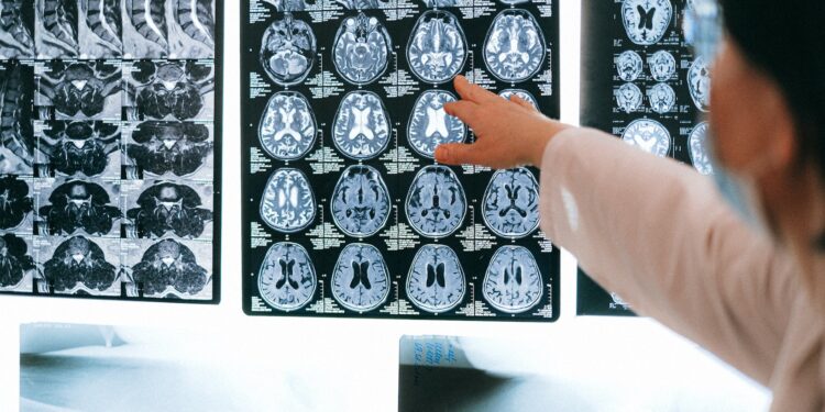
The lateral projection of a vertebra offers a side view of the bone, providing valuable information about its shape, size, and alignment. By examining this projection, healthcare professionals and anatomists can identify any fractures, deformities, or pathologies present in the vertebra. This knowledge plays a crucial role in diagnosing conditions such as spinal fractures, degenerative disc disease, and spinal tumors. Understanding the lateral projection of a vertebra is essential for accurate diagnosis and effective treatment planning.
Techniques for Identifying a Lateral Projection
When it comes to identifying a lateral projection of a vertebra, healthcare professionals rely on various techniques to ensure accurate diagnosis and treatment. These techniques utilize imaging modalities such as X-rays and CT scans to provide a clear side view of the vertebrae. Let’s explore some of the methods commonly used in identifying a lateral projection:
- X-ray Imaging: X-rays are a widely used diagnostic tool for identifying lateral projections of vertebrae. In this technique, a small amount of radiation is directed through the body, creating an image on a specialized film or digital sensor. By analyzing the resulting X-ray image, healthcare professionals can identify any fractures, deformities, or pathologies in the vertebrae. X-ray imaging is particularly helpful in detecting spinal conditions such as scoliosis or compression fractures.
- CT Scans: Computed Tomography (CT) scans are another valuable technique for identifying lateral projections of vertebrae. CT scans use a series of X-ray images taken from different angles to create detailed cross-sectional images of the spine. This imaging technique provides a more comprehensive view of the vertebrae, allowing healthcare professionals to accurately identify any abnormalities. CT scans are especially useful in diagnosing conditions like herniated discs or spinal tumors.
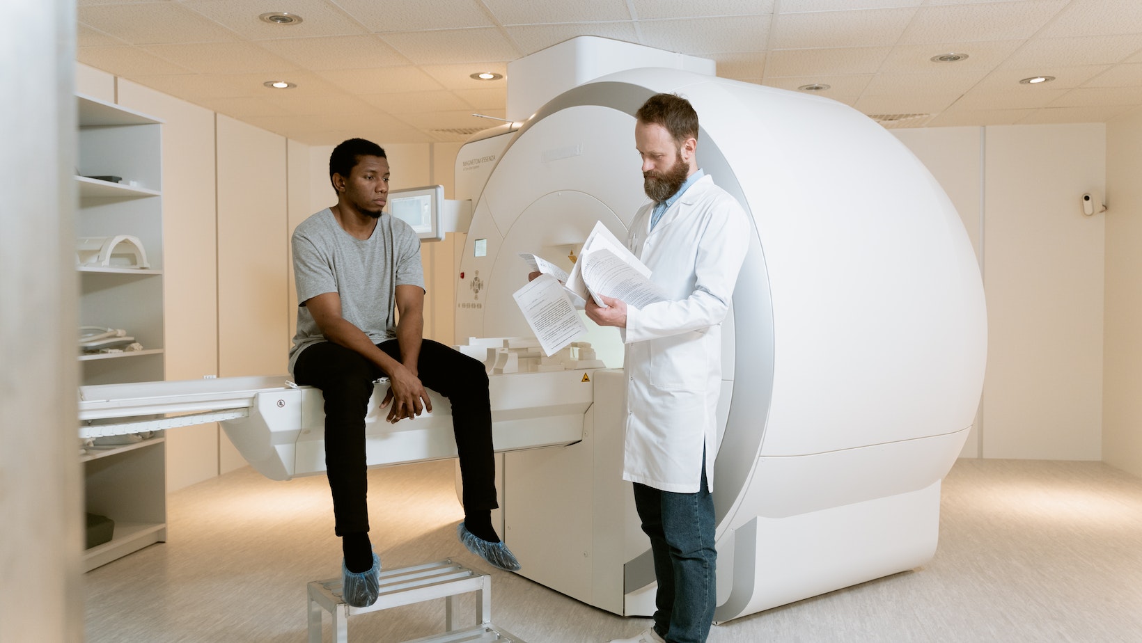
- MRI: Magnetic Resonance Imaging (MRI) is a non-invasive technique that uses magnetic fields and radio waves to create detailed images of the vertebrae. While MRI scans are not commonly used for identifying lateral projections, they are highly effective in visualizing soft tissues, such as spinal discs, ligaments, and nerves. By obtaining a clear picture of the surrounding structures, healthcare professionals can better understand the context of a lateral projection and make informed decisions about treatment.

Identify A Lateral Projection Of A Vertebra.
When it comes to identifying a lateral projection of a vertebra, healthcare professionals may encounter certain challenges. These challenges can make the process more complex and require additional expertise. Here are some of the common challenges faced in identifying a lateral projection of a vertebra:
- Obtaining the correct positioning: One of the main challenges is ensuring that the patient is positioned correctly for the imaging technique being used. This is crucial to obtain accurate lateral projections. Improper positioning can result in distorted or unclear images, making it difficult to identify any abnormalities or pathologies.
- Overlapping structures: In some cases, the lateral projection may be obstructed by overlapping anatomical structures. This can include nearby vertebrae, ribs, or soft tissues. Distinguishing the lateral projection from these overlapping structures requires a keen eye and experience in interpreting the images.
- Variations in patient anatomy: Every patient’s anatomy is unique, and this can pose a challenge in identifying a lateral projection. Variations in spine curvature, bone density, or even patient movement can affect the clarity and visibility of the lateral projection. Healthcare professionals need to account for these variations and adapt their techniques accordingly.
- Limited imaging resolution: The resolution of imaging techniques can vary, and in some cases, the lateral projection may not be as clear as desired. This can be due to limitations in X-ray machines, CT scans, or MRI equipment. Healthcare professionals need to rely on their expertise and utilize any available tools or software to enhance the clarity of the lateral projection.
- Interpreting the findings: Even when a lateral projection is successfully obtained, interpreting the findings accurately can be challenging. Identifying fractures, deformities, or pathologies within the vertebrae requires a comprehensive understanding of spinal anatomy and pathology. It’s important to consider the clinical context and correlate the findings with the patient’s symptoms and medical history.
Identifying a lateral projection of a vertebra can present challenges such as obtaining correct positioning, dealing with overlapping structures, variations in patient anatomy, limited imaging resolution, and interpreting the findings accurately. Overcoming these challenges requires expertise, experience, and a thorough understanding of spinal imaging techniques. By addressing these challenges, healthcare professionals can ensure accurate diagnosis and effective treatment planning for various spinal conditions.


